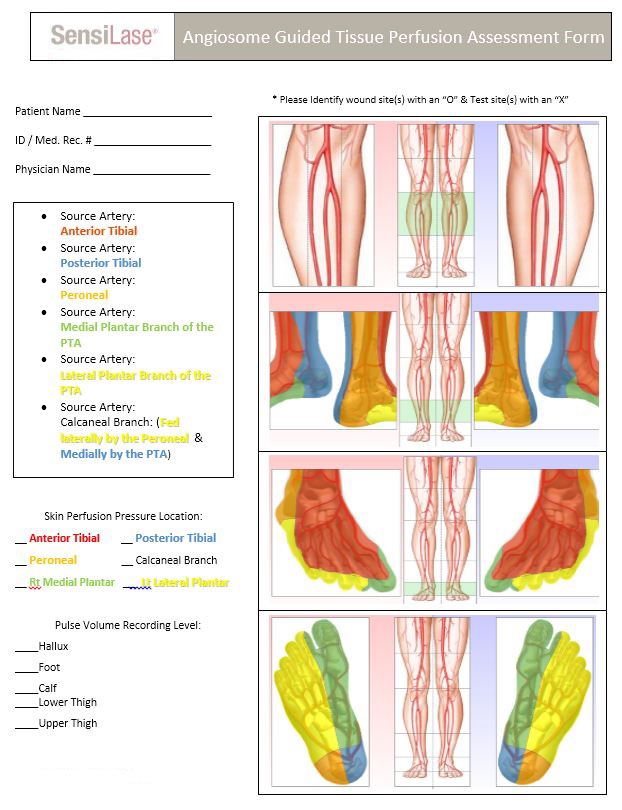
Angiosome Guided Testing
Targeted perfusion assessment based on arterial territories. The six angiosomes of the foot and ankle allow clinicians to evaluate specific vascular supply zones for precise diagnosis and treatment planning.
An angiosome is a three-dimensional block of tissue (including skin, subcutaneous tissue, fascia, muscle, and bone) supplied by a specific source artery. The angiosome concept, introduced by Taylor and Palmer, divides the foot and ankle into six distinct vascular territories fed by three main arteries below the knee.
Understanding angiosome anatomy is crucial for wound care, amputation planning, and revascularization decisions because different areas of the foot receive blood supply from different arterial sources.
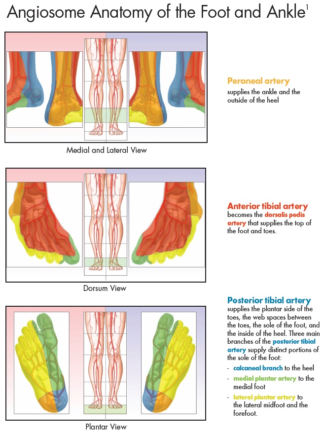
Peroneal Artery
Supplies the lateral ankle and outside of the heel.
Anterior Tibial Artery
Becomes the dorsalis pedis artery that supplies the top of the foot and toes, as well as the anterior shin.
Posterior Tibial Artery
Supplies the plantar side of the toes, the web spaces between the toes, the sole of the foot, and the inside of the heel.
Three main branches supply distinct portions of the sole:
- Calcaneal branch - to the medial heel
- Medial plantar artery - to the medial foot (instep)
- Lateral plantar artery - to the lateral midfoot and forefoot
Angiosome-guided testing is particularly important for diabetic patients because:
- The posterior tibial artery is most likely to occlude in diabetics - and this artery supplies the plantar foot where most diabetic ulcers occur
- TcPO2 cannot test plantar foot due to callused/thick skin - SPP has no such limitation
- Different parts of the foot may have very different perfusion levels based on which arteries are affected
- Revascularization planning benefits from knowing which specific angiosome requires improved blood supply
| Source Artery | Angiosome Region | SPP Test Location |
|---|---|---|
| Anterior Tibial | Dorsum of foot and ankle | Foot - Dorsal |
| Posterior Tibial | Medial heel | Calcaneal Branch |
| Medial sole/instep | Medial Plantar | |
| Lateral midfoot/forefoot | Lateral Plantar | |
| Peroneal | Lateral ankle and heel | Lateral Ankle / Calcaneal |
Wound Assessment
By testing the specific angiosome where a wound is located, clinicians can determine whether adequate perfusion exists for healing without intervention.
SPP measurement can disclose isolated obstruction in 1 or 2 of the 3 branches of the popliteal artery between the knee and the ankle.
Revascularization Planning
Angiosome-directed revascularization targets the specific artery feeding the wound location for optimal healing outcomes.
Post-revascularization, SPP can immediately confirm improved perfusion - unlike TcPO2 which requires 3-4 weeks to show improvement due to edema.
PAD-IQ Touch Screen Interface
PAD-IQ's color-coded touch screen interface makes angiosome-guided testing intuitive. Clinicians can easily select the appropriate test locations based on the angiosome anatomy displayed on screen, ensuring accurate and targeted perfusion assessment.